Plenary Speakers
A hugely impressive line-up of Plenary Speakers has been confirmed for mmc2015, and in recognition of their work, several will be receiving RMS Honorary Fellowships during the event. This is the Royal Microscopical Society's highest award.
Confirmed Plenary Speakers
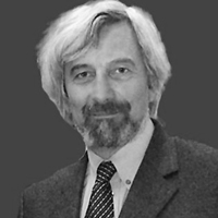
 Dirk van Dyck graduated from the University of Antwerp, Belgium in 1976 and has spent his career there since. He is now a Professor in Physics and Honorary Vice-Rector for research at the University. He teaches theoretical physics, image processing and information theory. Dirk Van Dyck was co-director of the EMAT centre for electron microscopy at the University of Antwerpand Director of the Vision lab, he remains involved in these as an Emeritus professor. He is known for his work in Electron Microscopy and microtomography and was a co-developer of the first table-top X-ray microtomograph that was commercialised by the spin-off company Skyscan, now Bruker MicroCT. Dirk Van Dyck has published approximately 300 scientific papers and several books. He has been promotor of 30 PhD Theses and holds 4 patents. In 2002 Dirk Van Dyck received the prestigious Francqui Chair from the University of Leuven, and in 2008 was presented with an Honorary Doctorship from the University of Lima, recognising his work and noting him as one of the most outstanding European experts in the area of Electron Microscopy.
Monday 29 June at 17:45 - Atomic resolution tomography and dynamics of nano-objects
 Dr Haider is co-founder and leader of CEOS GmbH, which has pioneered the development of spherical and chromatic aberration correctors, and produces the majority of correctors currently installed in transmission electron microscopes.
Dr Haider studied physics in Kiel and Darmstadt and received his PhD in 1987 in Darmstadt with an experimental work which was carried out at the Europaen Molecular Biology Laboratory (EMBL.) In 1989 he became a Group Leader withing the Physical Instrumentation Program at the EMBL. In 1992 he started a project to set up a Cs-corrector for a 200kV TEM together with Prof Rose in Darmstadt and Prof Urban in Juelich. In 1996 he founded CEOS GmbH in Heidelberg with Joachim Zach.Dr Haider is one of the pioneers of technology that overcame the 60 year old problem of spherical aberration in the electron microscope and has now revolutionised the performance of the TEM. His work has also encompassed monochromators and detector technologies in STEM.
In 2011 he was awarded the Wolf Prize in Physics, and at mmc2015 he will be awarded with an RMS Honorary Fellowship.
Thursday 2 July at 11:00 - Advanced instrumentation for high resolution EM
 Colin Humphreys is distinguished for his outstanding contributions to electron microscopy of materials. In his early work he developed theories of inelastic scattering and absorption of electrons passing through materials, of channelling patterns, and of SEM image contrast. He used electron diffraction scattering factors from critical voltage experiments to obtain bonding charge density maps for several elemental structures, and electron energy loss spectroscopy to demonstrate covalent bonding in transition metal aluminides. In GeSi/Si heterostructures, he discovered a novel source of misfit dislocations. Since 2000 he has carried out extensive studies of InGaN quantum wells used in LEDs which are remarkable for emitting bright light in spite of high dislocation densities. Combining electron microscopy and uniquely 3-D atom probe studies (with Cerezo), he showed that indium is distributed randomly (not clustered as previously thought), and that steps observed at interfaces provide a mechanism for carrier trapping. This work represents a significant advance in understanding the properties of these important energy saving devices. Colin is recognised as a world leader in this field. His InGaN work has attracted much industrial interest, resulting in several collaborations with industry aimed at developing the technology. Colin has also done outstanding work in promoting science, particularly in schools. He was knighted in 2010 for Services to Science.
Wednesday 1 July at 08:45 - Electron Microscopy: a Key Technique to help Save Energy, Purify Water and Improve our Health
 Jackie Hunter joined BBSRC as Chief Executive in October 2013. Jackie has over thirty years of experience in the bioscience research sector, working across academia and industry and playing a key role in innovative collaborations and partnerships. She holds a personal chair from St George’s Hospital Medical School, which was awarded in recognition of her contribution to bioscience research.
Jackie Hunter gained her first degree in Physiology and Psychology at the University of London followed by her PhD which was carried out at the Zoological Society of London. She undertook a Wellcome Trust post-doctoral research fellowship at St George’s Hospital Medical School before taking a role in the pharmaceutical sector in 1983.
During her career in industry, as well as leading neurology and gastrointestinal drug discovery and development, Jackie also spent several years developing and leading GSK's external science engagement strategy. In this role she played a central part in fostering effective and innovative collaborations and partnerships between university and institute research groups and the company.
Jackie is a current member of the Council of the University of Hertfordshire and previously served on the Council of Royal Holloway University of London as well as the governing body of the Babraham Institute. From 2004, Jackie was a member of BBSRC Council and BBSRC Strategy Board. She is a fellow of the British Pharmacological Society.
She founded OI Pharma Partners in 2010 to support the life science sector in harnessing the power of open innovation. Open innovation allows public and private organisations to use a range of collaborative models to find the best ways to bring ideas to fruition. Jackie was awarded a CBE in the Queen's Birthday Honours list for Services to the Pharmaceutical Industry as well as the Women of Achievement in Science, Engineering and Technology (SET) awards in the category SET Discovery, Innovation and Entrepreneurship in 2010.
Monday 29 June at 17:00 - The Evolution Of Biological Microscopy - From Form To Function
 Petra Schwille is Director of the Research Department for Cellular and Molecular Biophysics at the Max Planck Institute for Biochemistry in Germany. Petra studied physics and then began to move into Biophysics as a PhD student, taking her to the Max Planck Institute for Biophysical Chemistry. She then worked at the Department of Applied and Engineering Physics at Cornell University, returning to Max-Planck in 1999 to take up the position of Junior Group Leader for Experimental Biophysics. In 2002 she became a Professor of Biophysics, teaching at Dresden University of Technology and then took up her current role in 2012. Petra’s research seeks to understand living systems at their most fundamental scale of interacting molecules. Her work has significantly advanced the development of Fluorescence Cross Correlation Spectroscopy. She has been recognised for her work via an extensive list of awards and Honorary positions, including the Braunschweig Research Prize, the Suffrage Science Award and elected membership to the National Academy of Science and Engineering and Berlin-Brandenburg Academy of Sciences and Humanities.
Tuesday 30 June at 08:45 - Bottom-up synthetic biology – a new tool for understanding the cell on a molecular level
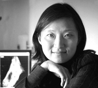
 Xiaowei Zhuang is a Professor of Chemistry and Chemical Biology and a Professor of Physics at Harvard University, and an investigator of the Howard Hughes Medical Institute. She is a biophysicist recognized for her work in the development and application of advanced optical imaging techniques for the studies of biological systems. In particular, she and coworkers invented a super-resolution fluorescence imaging method, Stochastic Optical Reconstruction Microscopy (STORM), which breaks the diffraction limit. Her laboratory continues to push the envelope of super-resolution imaging, and her recent innovations in optical methods and fluorescent probes allowed the resolution of STORM to reach a few nanometers. The ultrahigh resolution provided by STORM is transforming biomedical research and has enabled discoveries of novel cellular structures. Zhuang and coworkers demonstrated the applicability of STORM to imaging a wide variety of biological systems ranging from single-cell organisms to complex tissues, and discovered novel super-molecular structures in the cytoskeleton and chromosomes. Zhuang and her lab members have also developed and applied single-molecule approaches to investigate the structure, dynamics and function of biomolecules, with emphasis on how proteins and nucleic acids interact and how protein-nucleic acid complexes function.
Thursday 2 July at 16:15 - Illuminating biology at the nanoscale with super-resolution imaging


